
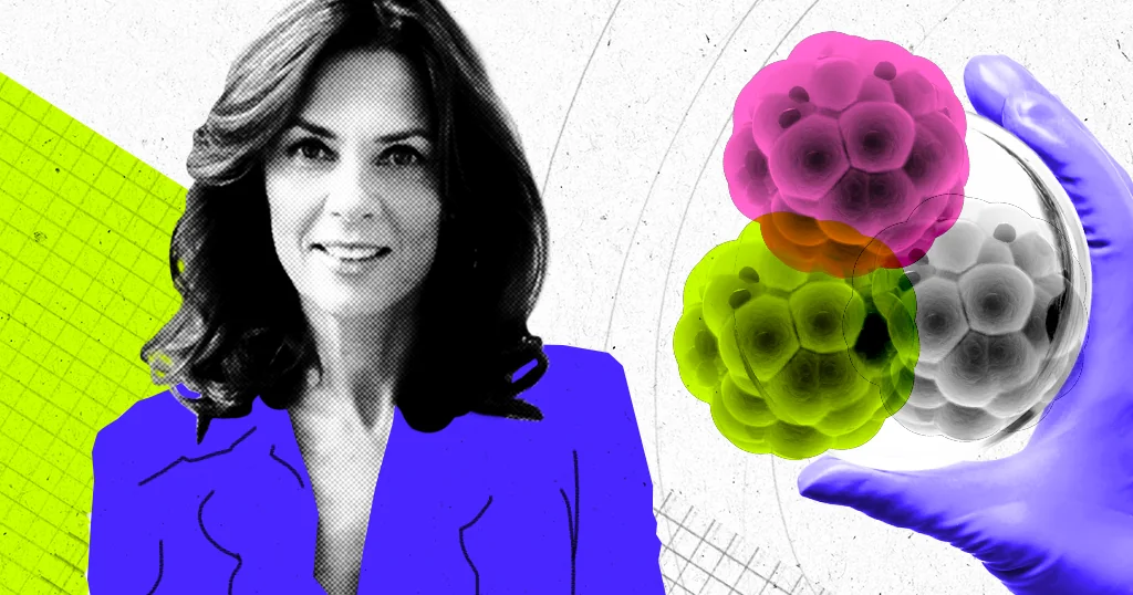
Magdalena Zernicka-Goetz is the Bren Professor of Biology and Biological Engineering at the California Institute of Technology and a Professor of Stem Cell Biology and Development at the University of Cambridge.
In this interview, we talk about recent breakthroughs in technologies that let us use stem cells to make embryo-like structures with a brain and even a beating heart in a dish. We look at how these ‘synthetic’ embryos are assembled and the extent to which they resemble natural embryos that develop from fertilized eggs. She also describes how they may help us learn why pregnancies fail, how to build organs from scratch, and even how to revitalize aging bodies. But first, she shares the central insight that has enabled us to keep these embryo models growing in a dish longer than ever before: the cells that will form the body cannot do it by themselves.
What is a synthetic embryo, and what can it be used for?
To begin, can you explain what a synthetic embryo is?
MAGDALENA ZERNICKA-GOETZ: To be honest, I don’t really like that phrase. It’s misleading, because people will ask: what is it made from?
But we use it because it is a shorthand for saying that we have created an embryo-like structure from building blocks. In our lab, we use three kinds of building blocks. One building block represents the stem cell for every kind of cell that will make up our adult body. It is called the embryonic stem cell. The other two building blocks are stem cells for the so-called extraembryonic structures. One of them is well known: the placenta. This is the part that links the baby to the mother’s body, through which the baby is nourished. The second extraembryonic structure is less well known, and it is called the yolk sac. That is a sort of sac in which the embryo grows.
Broadly, what are some of the things we might want to do with synthetic embryo models?
So, for instance, we have shown that these models can be used to understand the role of specific genes that are essential for certain stages of development. We know, for example, that there is a gene that matters for brain and eye development. But we do not know exactly how it works from real mouse embryo models, because we cannot track the entire process from start to finish so precisely. So now you can use embryonic stem cells, remove that gene, and learn more about the stage of development this gene matters for and what it does. You can also delete these genes at different time points and observe the effects.
It will not be able to grow and develop as we do, but it can give us important insight into the fragments of life that at this moment are a total mystery.
We can also examine the role of a particular environment or specific metabolites. For example, pregnant women are told to take folic acid because it supports neural development. But at which exact stage is it important, what does it actually do?
Is there a chance to better understand why so many pregnancies end very early, given these models replicate the same early stages of development?
Yes, absolutely. It is very important to understand that most pregnancies fail at the stage when we do not even know we are pregnant. The first two weeks of development are very delicate because there are major milestones that must be reached at the right time.
First, we need to generate stem cells for the three tissues I mentioned, two extraembryonic and one embryonic. We have to make them correctly, and then those tissues need to interact with one another. But timing matters too. You cannot lengthen pregnancy to, say, 15 months. This shows that specific milestones have to be reached at specific time points.
Only one stem cell type really builds the body, but the other two are guiding forces, a little like a mother and father.
So when these developmental milestones are not happening properly, or they are delayed, or happen too early, embryos are aborted. Or when communication between those three kinds of cells is somehow abnormal, or not happening at all, again, embryos become aborted. That is why so many pregnancies fail. So now, with these models, we are able to investigate how we can protect the baby within the body of the mother. That is the hope and that is a very important motivation for me.
I do want to emphasize, though, that right now we are discussing synthetic mouse embryo models. But clearly, this is a kind of prototype for creating human three-dimensional embryo models, but even then it would not truly be a human embryo. It will not be able to grow and develop as we do, but it can give us important insight into the fragments of life that at this moment are a total mystery.
So where are we with human synthetic embryo models or even culturing in vitro human embryos?
So, human embryo models are not there yet. To my knowledge, there is not yet a complete embryo-like structure made from human stem cells. When we began building stem cell-derived mouse embryo models, many people asked why we were not doing it with human stem cells, and I am sure that many of my colleagues are trying to build a similar model using human stem cells. But it is not straightforward. First of all, human stem cells and mouse stem cells do not develop in the same way. They need different conditions to be maintained in culture. To really make sure that we know how to do it, the mouse model will be a prototype.
Still, many people, including us, use human stem cells in culture to build three-dimensional tissues or fragments of embryos. We use them to understand, for example, how the amniotic cavity (the closed sac that contains the amniotic fluid), forms. Would we be able to correct its development when it goes wrong?
But it is only a fragment of the human embryo, a model at the early stages of implantation in the uterine wall. Right now, we can culture human embryos only until so-called day 14, this is the limit where we cannot pass.
Creating embryo-like structures in the lab
That is fascinating. So, how do you create the mouse synthetic embryo?
The way we build these synthetic embryo models in our lab is rather unique. We developed this approach by understanding how the embryo builds itself in natural life, and we use what the embryo teaches us to imitate that process in the lab in a petri dish.
So we use the three kinds of stem cells. We try to combine them in the correct proportions, create the right environment so that the three kinds of cells, and the cells that will come from them, are happy and want to communicate with each other.
That is what matters most: using three kinds of cells, not one, because development normally happens through interactions among three cell types. Only one stem cell type really builds the body, but the other two act as guiding forces, a bit like mother and father.
I have never put it that way before, but you could see it like this because these two other cell types give instructions and signaling information, and they also create a kind of home for the embryo to be nourished in there properly.
Let us step back a little. This field has advanced a lot over the last few years. Can you tell me what the truly important milestones have been in making progress toward building this embryo model overall?
I have to state two well known facts. First, embryonic stem cells can be kept in culture and can keep multiplying in culture indefinitely. That was Martin Evans's discovery, and he won the Nobel Prize for it. We knew that if you took a few of those cells and put them together with an embryo, they could contribute to adult tissues later on in developing organs too.
So we knew that stem cells have this remarkable potential. But what we did not know, and what became a breakthrough about 10 years ago, was whether we could build embryos entirely from those cells, without the host embryo. It was not a sudden event, of course; it happened step by step. But the way we learned to do it was by first watching how the embryo does it in real time in culture.
There is a very early stage of development called implantation, and we know very little about it, especially in humans. The first few days before that stage are fairly well understood. The three cell types I mentioned arise during those first few days of development from the embryo’s starting point in the lab.
[These] models are important not only for understanding embryogenesis, but also for understanding the origin of particular tissues that build our adult organs. We are trying to identify the basic rules that must be met in each case here.
Once these three cell types are formed, they begin to communicate with one another. But how they communicate was not well known, because this is the time when the embryo invades the mother’s body in the process called implantation. We could not mimic this process in vitro, so we could not observe it. So our first step was to develop a way to culture real embryos, mouse and human, through that stage in the lab there.
As soon as we were able to do that, we could follow the cells, label them, and track them to identify the time when they multiply and interact with one another. When we followed those events, we realized that now we knew enough to mimic these events with stem cells representing the three tissues in a controlled laboratory setting.
It was a journey, and the first, most important milestone was figuring out how the embryo does it. In particular, we realized that the embryo takes instructions from the two extraembryonic tissues. So far, we have built five models by adding different combinations of extraembryonic cells to the embryonic ones. The first model was published in 2014, and the last model was published.
Tell me about this next step. What has been achieved with this new model in terms of how far the embryos progress and what you can see in them? And how do they look compared with a fertilized egg that develops into an embryo?
The last model now develops until the moment when the head, heart and somites, segments along the body axes, form. This is incredible, because we were not sure whether these embryo like structures would be good enough to reach these milestones. All the progenitors of the brain are there, and the heart structure beats and pumps blood.
The lessons from the early embryo can also teach us how to rejuvenate tissues, because embryonic tissues are young tissues.
So how similar are they to natural embryos? They are very similar, but not identical. This is very interesting, because then you can follow the development of the models that are nearly identical, and those that are not, to understand the basic principles that we have to fulfill to make a particular type of tissue or organ perfect.
That is why those models are not only important for us to understand embryogenesis, but also important for understanding the origin of particular tissues that build our adult organs. We are trying to identify the basic rules that must be met for these events to be accomplished properly. You can start to work out what is going on, and since you are letting the embryo build itself, you can work out the mechanisms of that process and when they go wrong.
Where synthetic embryos might lead
Tell me a little more about what you personally want to do with these models. Are there particular questions or challenges that you want to tackle directly?
My main interests are twofold. First, I want to understand how life is created. So I use this model to try to really understand this mysterious stage of life when cells communicate with each other for the first time to build something as complex as we are. But this is also the time when most pregnancies fail. If we can understand this, we may in the future be able to help prevent those failures. That is our hope for now.
It is a little like how to build a house, right? You do not rely on the building blocks to sort themselves out alone.
The lessons from the early embryo can also teach us how to rejuvenate tissues, because embryonic tissues are young tissues. So it teaches us about building our organs and building tissues. Hopefully the knowledge from these studies, step by step, will be used for transplantation of organs or repairing organs in our adult bodies when they fail.
Are there existing roadblocks, either technical or in our scientific understanding, that are holding back the development and use of these models?
Yeah, there are, mainly around the technology of creating the embryo like structures. When we put these three types of stem cells together, we rely on the forces between them to create the proper embryo. Sometimes that goes well, sometimes that does not go well. We see this variability of structures. So we will have to develop tools to control these events better.
For example, at this conference I am currently attending, I spent time discussing optogenetics with a colleague. Using light, he can stimulate particular responses of the cell. So can we use these optogenetic approaches to help guide the process of self organization?
To guide the process in what way?
To design particular events. For instance, when we imagine making tissues and organs that can replace damaged ones, doing that efficiently would require us to know how to engineer them. It is somewhat like constructing a house, right? You do not expect the building blocks to arrange themselves. Or, if a building were less than perfect, that would be unacceptable. We would want to direct the building process from the start so there is quality control as well.
So, we are not yet able to act as engineers or architects. Instead, we are trying to create an environment in which the embryo can build itself, and to understand that process, observe it, and help it or perturb it. But we are not yet doing tissue engineering. Tissue engineering is very, very important, and it will be the future of organ replacement. So many patients are waiting for liver transplants, or other organs that are failing, and this is truly tragic. If we can create and repair those organs using the knowledge that comes from our studies, it will be absolutely incredible. What we do, and what many of my colleagues do—so-called bioengineering of tissue—is where it is going in the future, at least.